Skip to content
Victoria Aesthetics Wellness Centre

LASER CENTRE · VICTORIA DEEP PIGMENT PRECISION™

Nevus of Ota, Hori & Dermal Pigmentation Treatment in Seychelles

Not all pigment sits on the surface.

Blue-grey or deeper facial pigmentation can come from pigment within the dermis rather than the epidermis. Victoria Deep Pigment Precision™ combines physician-led diagnosis, pigment-depth assessment and selected laser strategies for Nevus of Ota, Hori-type pigmentation (ABNOM) and other appropriate dermal pigment patterns.

  • Physician-supervised
  • Diagnosis before laser
  • Skin-of-colour aware

Assessment SCR 900 · deductible from your treatment plan

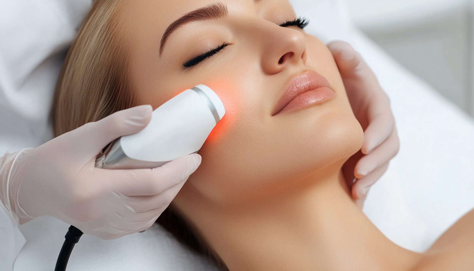
Physician-supervised facial pigment laser treatment at Victoria Aesthetics, Seychelles

FIRST STEPDeep Pigment Mapping™

STRATEGYMulti-wavelength · 1064 nm

SUPERVISIONDr. Eric Licea, MD

  • CONCERNS

    Ota · Hori

    And selected dermal or mixed-depth facial pigmentation

  • TREATMENT

    A course

    Staged sessions with biological review between them

  • NEVUS OF OTA

    SCR 5,500

    Per session, for a clinically mapped defined region

  • ASSESSMENT

    SCR 900

    Deductible from your treatment plan

Close-up of facial skin assessed for pigment colour and distribution
Colour≠Diagnosis

BLUE-GREY DOES NOT AUTOMATICALLY MEAN NEVUS OF OTA

Blue-grey pigment has more than one possible diagnosis.

Colour gives clues about pigment depth, but appearance alone does not establish the diagnosis. A blue-grey, slate-grey or brown-grey facial pattern may represent different pigmentary disorders that need different treatment strategies.

  • Nevus of Ota (oculodermal melanocytosis)
  • Hori-type pigmentation / ABNOM
  • Dermal or mixed post-inflammatory hyperpigmentation
  • Melasma with deeper or mixed pigment components
  • Other dermal melanocytoses or pigmented lesions that need medical differentiation

Same colour does not mean same condition.

THE COLOUR IS A CLUE

The depth changes the plan.

WHY DEEP PIGMENT LOOKS BLUE OR GREY

Why can deeper pigment look blue-grey?

Melanin located deeper in the skin can look grey, blue-grey or slate-coloured rather than simply brown. That visual effect reflects how light interacts with pigment at different depths.

Depth matters because a treatment designed for superficial, epidermal pigment may not be the right strategy for dermal melanocytes or dermal melanophages.

Select a depth to see where the pigment sits and how it tends to look. Simplified and not to scale: colour alone cannot measure depth precisely.

DERMAL PIGMENT

Pigment-producing cells or pigment sit within the dermis. Typical of Nevus of Ota and Hori-type pigmentation, and usually treated as a staged course.

The colour is a clue. The depth changes the plan.

TWO CONDITIONS, NEVER SYNONYMS

Nevus of Ota and Hori-type pigmentation.

Both are forms of dermal pigmentation. They differ in when they appear, where they sit on the face and how they are planned, which is why we assess, plan and price them separately.

UNILATERAL · OFTEN PRESENT EARLY IN LIFE

Nevus of Ota

A dermal melanocytosis classically producing blue-grey pigmentation in the distribution of branches of the trigeminal nerve, often around the eye, temple, forehead, cheek or nose. It is commonly one-sided, although presentations vary.

Look
Blue-grey to slate-grey
Laser context
Q-switched pigment lasers have the longest track record; picosecond 755 nm and 1064 nm approaches also have published evidence.
Course
A series rather than a single session; no universal session number.

When pigment extends beyond the skin. Nevus of Ota can involve the eye. A cosmetic laser session treats selected skin pigment; it does not assess or treat ocular involvement. Tell us about any pigment in the white of the eye, visual symptoms or previous eye follow-up, as an ophthalmic assessment may be advised.

BILATERAL · ACQUIRED IN ADULTHOOD

Hori-type pigmentation / ABNOM

Acquired bilateral nevus of Ota-like macules, also called Hori's nevus: an acquired dermal melanocytosis that typically shows as bilateral blue-grey to grey-brown macules, commonly over the cheekbones. It can resemble melasma, or coexist with it.

Look
Speckled grey-brown to blue-grey, both cheeks
Laser context
Laser can produce meaningful improvement; protocols vary and post-inflammatory hyperpigmentation is a significant consideration.
Course
Improvement is usually progressive over several sessions.

Why the diagnosis matters. Hori-type pigmentation and melasma have different biology. Treating every bilateral pattern as melasma, or every grey-brown patch as ABNOM, can lead to the wrong strategy.

TELLING THEM APART

Deep pigment, Hori or melasma?

More than one condition can coexist. Hori-type pigmentation with melasma matters clinically, because treatment response and the risk of post-inflammatory pigmentation can differ.

PatternUsually looks likePathway
Nevus of OtaOften blue-grey; classically one-sided in a characteristic distribution; may involve the eye.Deep Pigment Precision™
Hori / ABNOMAcquired, bilateral, discrete or mottled grey-brown to blue-grey macules over the cheekbones.Deep Pigment Precision™
MelasmaUsually broader, patch-like and often symmetrical; chronic, recurrent and multifactorial.Victoria Melasma Control™
Post-inflammatory hyperpigmentationAppears after inflammation or injury and follows where that event happened.Victoria PIH Recovery™
Sun spots & frecklesDiscrete, sun-related brown spots sitting near the surface.Victoria Sun & Spot Precision™

DIAGNOSTIC ROUTER

What pattern does your pigmentation resemble?

A pattern is a starting point for a conversation, never a self-diagnosis. The final answer always comes from the clinical assessment.

  • One-sided blue or slate-grey patch

    Consider a Nevus of Ota pattern

    Deep pigment assessment

  • Speckled grey-brown macules on both cheekbones

    Consider Hori / ABNOM

    Deep pigment assessment

  • Broad symmetrical brown or grey-brown patches

    Consider melasma or a mixed pattern

    Melasma assessment

  • Pigment that followed acne, irritation or injury

    Consider post-inflammatory hyperpigmentation

    PIH assessment

  • Discrete sun-related brown spots

    Consider lentigines or freckles

    Sun & spot assessment

  • New, changing, irregular or symptomatic pigment

    Medical evaluation comes first

    Before any cosmetic laser

The website can recognise a pattern. Only the clinical assessment can establish the pathway.

VICTORIA DEEP PIGMENT MAPPING™

The deeper the pigment, the more important the diagnosis.

Before any laser plan, we evaluate the pattern, colour, distribution, likely pigment compartment, skin type, coexisting pigment disorders and how your skin has responded to previous treatment.

  • Map one-sided versus bilateral distribution
  • Assess the eye area, cheekbones, temple, forehead, cheek and nose
  • Look for features of Nevus of Ota, Hori / ABNOM, melasma, PIH or mixed disease
  • Assess skin phototype, recent tanning, PIH history and previous laser response
  • Record a baseline with standardised photography
  • Decide whether an eye or dermatology assessment is indicated
  • Decide whether laser is appropriate now, should be staged or should wait

We do not choose the wavelength until we understand the pigment.

Facial pigment distribution assessed before treatment planning

THE TECHNOLOGY

Why is 1064 nm often discussed for deeper pigment?

The longer 1064 nm wavelength reaches deeper and is absorbed less by the melanin at the surface than shorter, visible wavelengths. That is one reason it has an established role in selected dermal pigment disorders, including Nevus of Ota. It is still a tool, not a guarantee.

  • 1064nm

    Nd:YAG

    Strong historical and contemporary evidence for selected dermal pigment, including in darker skin types, with a gentler interaction with surface melanin.

  • 755nm

    Alexandrite (picosecond)

    Published evidence for Nevus of Ota and Hori / ABNOM. Effective in studies, although post-inflammatory hyperpigmentation and hypopigmentation remain possible.

  • 532nm

    Visible green

    May have a role for selected superficial or mixed components in combination protocols. Not a default wavelength for deep pigment.

Wavelength alone does not decide safety or success. Pulse duration, energy, spot size, treatment endpoint, interval, the biology of the lesion, your skin type and clinical judgement all matter. 1064 nm is a tool. Diagnosis is the strategy.

YOUR DEEP PIGMENT JOURNEY

A long-term plan without a long-term promise.

Deep pigment is usually a course, not a one-session event. The target sits deeper, and over-aggressive single treatments raise the risk of side effects, so we work in stages and let your skin show its response before the next step.

  1. PHASE 01 · IDENTIFY & MAP

    Diagnose & baseline

    Deep Pigment Mapping™, standardised photography, eye or medical referral when indicated, and the treatment strategy.

  2. PHASE 02 · TARGET

    Initial targeting

    The first treatment phase, with the wavelength and parameters selected for your diagnosis and skin.

  3. PHASE 03 · REVIEW

    Biological review

    Time for the skin to show fading, any post-inflammatory pigment, lightening or mixed behaviour before anything is escalated.

  4. PHASE 04 · CORRECT

    Progressive correction

    Treatment is repeated or modified only when the previous response supports it.

  5. PHASE 05 · FOLLOW

    Consolidate & follow up

    We look at residual pigment, stability and recurrence, and whether further treatment adds meaningful value.

Progressive clearance is not treatment failure. It is often the biology of deep pigment.

MELANIN-RICH SKIN · PIH-AWARE PLANNING

Treating deep pigment means respecting the melanin around it.

Many people with dermal pigmentation have melanin-rich skin. Laser energy has to reach the abnormal pigment while sparing the normal pigment above it as much as possible. That is why you will never read “safe for all skin types” on this page.

  • Post-inflammatory hyperpigmentation can occur after treatment
  • Hypopigmentation (lightening) can also occur and may persist
  • More energy is not automatically more effective
  • Recent tanning or sunburn can change whether you can be treated
  • Intervals matter: the skin needs time to show its response
  • Coexisting melasma may need its own control strategy
Melanin-rich skin, planned with conservative pigment-laser parameters
Conservative·Staged

AFTER LASER

What can my skin look like after treatment?

Deep pigment does not need to disappear straight away for treatment to be working. The response develops gradually, and we judge it at review, not in the mirror the next morning.

  1. IMMEDIATELY

    Redness, swelling, whitening or other temporary endpoints may appear, depending on wavelength and protocol.

  2. FOLLOWING DAYS

    Pigment may look temporarily darker; mild crusting or other surface changes can occur.

  3. FOLLOWING WEEKS

    Change develops gradually as the body clears fragmented pigment.

  4. AT REVIEW

    We compare photographs and assess fading, residual pigment and any coexisting melasma before deciding the next step.

Contact the clinic about unexpected blistering, significant pain, signs of infection, prolonged inflammation or any pigment change that worries you.

REALISTIC OUTCOMES

What can we realistically aim for?

Long-term behaviour depends on the diagnosis. Recurrence has been reported in Nevus of Ota even after substantial clearance, and Hori-type pigmentation can be complicated by melasma or new pigment changes. A good result is one that stays stable and healthy.

WE AIM FOR

Progress you can see, and measure.

  • Progressive visible reduction of correctly diagnosed dermal pigment
  • Less contrast between the affected area and surrounding skin
  • A staged plan that balances clearance with pigment safety
  • Objective photographic review, not memory alone
  • Separate management of coexisting melasma or PIH

WE DO NOT PROMISE

What no honest clinic can guarantee.

  • Complete clearance in every patient
  • A fixed number of sessions, or one-session removal
  • Zero post-inflammatory pigment or lightening
  • That the pigment can never return
  • That every blue-grey pattern should be lasered

OUR TREATMENT INTEGRITY PROMISE

If your pigment is not a laser-first condition, we will tell you.

If the pattern fits melasma, post-inflammatory pigmentation, an uncertain lesion or a condition that needs another medical evaluation, we redirect the plan before anything is sold.

The right laser on the wrong diagnosis is still the wrong treatment.

TREATMENT INVESTMENT

A treatment plan built around the diagnosis.

Nevus of Ota and Hori-type pigmentation are both dermal, but they rarely present in the same pattern or over the same surface area, so we price them separately. We sell a clinical pathway, not a guaranteed number of sessions.

NEVUS OF OTA

Targeted deep pigment treatment

For clinically diagnosed Nevus of Ota involving a defined facial region.

1 SESSION

SCR 5,500

  • Physician-supervised treatment of the mapped area
  • Individual laser parameter selection
  • Standardised clinical photography
  • Aftercare instructions and clinical review

INITIAL PHASE

3-SESSION PLAN

SCR 14,000

You save SCR 2,500

  • 3 physician-supervised sessions
  • Victoria Deep Pigment Mapping™
  • Progress photography and reassessment between stages
  • Parameters adjusted to your response

Three sessions are an initial treatment phase, not a guarantee of complete clearance. The total number varies with depth, extent, skin type and response.

Start my Nevus of Ota plan (opens the booking system in a new tab)

HORI / ABNOM

Bilateral deep pigment treatment

For clinically diagnosed Hori-type pigmentation, typically involving both cheekbone areas.

1 SESSION

SCR 7,500

  • Physician-supervised bilateral treatment
  • Individual laser parameter selection
  • Standardised clinical photography
  • Aftercare instructions and clinical review

INITIAL PHASE

3-SESSION PLAN

SCR 19,000

You save SCR 3,500

  • 3 physician-supervised bilateral sessions
  • Victoria Deep Pigment Mapping™
  • Evaluation for coexisting melasma or PIH
  • Parameters adjusted to your response

Hori-type pigmentation usually needs staged treatment. The plan is an initial correction phase followed by reassessment, not a promise that treatment ends after three sessions.

Start my Hori / ABNOM plan (opens the booking system in a new tab)

NOT SURE WHICH TYPE YOU HAVE?

Do not choose a treatment by colour alone.

Deep Pigment Mapping™ establishes whether your pigment fits Nevus of Ota, Hori / ABNOM, melasma, post-inflammatory pigmentation or something else before any treatment begins. The SCR 900 assessment is deductible from your plan.

Book my deep pigment assessment (opens the booking system in a new tab)

WHY VICTORIA AESTHETICS

Deep pigment needs more than a powerful laser.

Medical direction by Dr. Eric Licea, MD, with a Laser Centre that treats pigment as a diagnosis first and a wavelength second.

  • 01

    Diagnosis first

    Nevus of Ota, Hori / ABNOM, melasma and post-inflammatory pigmentation are not interchangeable.

  • 02

    Depth-aware planning

    The strategy reflects where the pigment is most likely located.

  • 03

    Multi-wavelength thinking

    The wavelength follows the diagnosis and the capability of the device.

  • 04

    Skin-of-colour aware

    The risks of darkening and lightening are discussed openly, not hidden.

  • 05

    Staged review

    Deep pigment is treated as a course, with biological reassessment between sessions.

  • 06

    One pigment ecosystem

    If another pathway fits better, such as melasma or PIH care, we route you there.

QUESTIONS

Frequently asked questions

If your question is about your own pigment, the deep pigment assessment is the place to answer it properly.

What is dermal pigmentation?

Pigment located within the dermis rather than only in the superficial epidermis. Different disorders can create dermal pigment, so the term describes location more than a single diagnosis.

What colour is dermal pigmentation?

It can look blue, blue-grey, slate-grey or grey-brown, although colour alone cannot reliably establish the diagnosis or the exact depth.

What is Nevus of Ota?

A dermal melanocytosis that commonly produces blue-grey pigmentation in a characteristic facial distribution, usually on one side, and can also involve the eye.

What is Hori's nevus, or ABNOM?

Acquired bilateral nevus of Ota-like macules: acquired dermal pigmented macules on both sides of the face, often over the cheekbones. They can resemble melasma or coexist with it.

Is Hori's nevus the same as melasma?

No. They have different biology, although their facial distribution can overlap and both can be present in the same person.

Can laser treat Nevus of Ota?

Yes, pigment lasers have an established role. Q-switched Nd:YAG has extensive historical evidence and picosecond systems also have published evidence. Treatment usually needs several sessions.

Can picosecond laser treat Hori / ABNOM?

Published evidence supports picosecond treatment, including 755 nm and 1064 nm approaches, but several sessions are typically needed and post-inflammatory hyperpigmentation remains an important risk.

Is 1064 nm always the best wavelength?

No. It is an important option for deeper pigment, but the best strategy depends on the diagnosis, your skin type, the lesion, the exact device and the treatment parameters.

How many sessions will I need?

There is no universal number. Published studies usually involve several treatments, and your course depends on the diagnosis, extent, depth, response and any side effects.

Can deep pigment be removed in one session?

That should not be expected. Some change may follow one treatment, but staged treatment is the norm for dermal melanocytosis.

Can the pigment come back?

Recurrence has been reported in Nevus of Ota, even after substantial clearance. Long-term behaviour depends on the diagnosis and your individual biology.

Is treatment safe for darker skin?

Treatment is possible in selected darker skin types, but post-inflammatory hyperpigmentation and hypopigmentation are recognised risks. Conservative, diagnosis-specific planning is essential.

What if I also have melasma?

Tell the clinical team. Coexisting melasma can change the response and may need its own control strategy rather than simply more laser energy.

Why might I need an eye assessment?

Nevus of Ota can involve the eye. Skin laser treatment does not replace an appropriate ophthalmic evaluation when eye involvement is present or suspected.

DERMAL PIGMENTATION · NEVUS OF OTA · HORI / ABNOM · MAHÉ

When pigment runs deeper, the plan must go deeper too.

  1. Identify
  2. Map
  3. Depth-assess
  4. Target
  5. Review

Victoria Aesthetic & Wellness Clinic · Bel Etang Building, Hermitage Road, Mont Fleuri, Mahé, Seychelles

DEEP PIGMENT ASSESSMENT

SCR 900

INCLUDES

  • Victoria Deep Pigment Mapping™
  • Pattern, distribution and skin-type assessment
  • Baseline photography
  • Pathway recommendation, or referral if needed
  • Deductible from your treatment plan

Medical disclaimer: Educational information only, not a substitute for an individual medical diagnosis. Blue-grey or deeper pigmentation has several possible causes. Laser suitability, wavelength, number of treatments, response and side effects vary. Post-inflammatory hyperpigmentation, hypopigmentation, incomplete response, recurrence, irritation, blistering, textural change and scarring are possible. Eye involvement: skin treatment of Nevus of Ota does not assess or treat the eye; an ophthalmic assessment may be advised when indicated.

Privacy and Cookies

This website uses cookies so that we can provide you with the best user experience possible. Cookie information is stored in your browser and performs functions such as recognising you when you return to our website and helping our team to understand which sections of the website you find most interesting and useful.

More information in Cookies polity